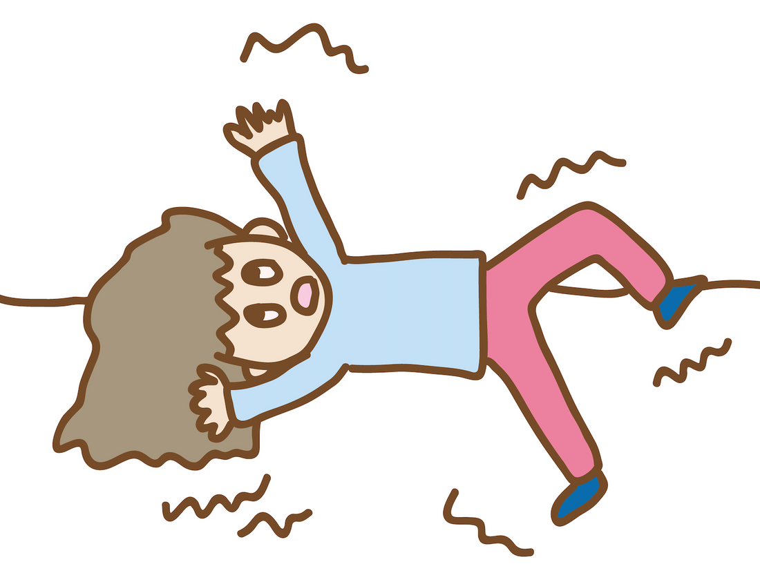
Myoclonus, one of the most commonly encountered involuntary movements, is characterized by sudden, brief, jerky, shock-like movements involving the extremities, face and/or trunk, without loss of consciousness. It concerns bursts of muscular activity.
There is a positive myoclonus (caused by abrupt muscle contractions) and there is negative myoclonus (sudden cessation of muscle contraction with silent periods of ongoing electromyographic activity).
Myoclonus can be classified into three groups:
• Cortical.
• Subcortical.
• Spinal myoclonus.
based on the presumed physiological mechanism underlying its generation.
Differentiation:
Cortical myoclonus is:
• Shock-like and focal, sometimes segmental.
• Appears on posture or movement (action).
• Can be irregular but also rhythmic.
• The stimulus is highly sensitive.
Subcortical myoclonus is:
• Less shock-like, and axial.
• Appears in rest.
• Tends to be periodic.
• The stimulus is not so sensitive.
Spinal myoclonus:
• Can be shock-like.
• Appears in rest.
• Seems to be periodic or rhythmic. (Rhythmic myoclonus can be mistaken for tremor).
• The stimulus can be sensitive.
The amplitude of myoclonus can vary considerably depending on the subtype. The lightning-like,
square-wave character of myoclonus helps in its differentiation from tremors.
Myoclonus does not necessarily represent a pathological phenomenon. Physiologic myoclonus
occurs during sleep transition or during sleep itself (hypnic jerks).
Cortical myoclonus
Cortical myoclonus generators are probably the most frequent neuroanatomical substrate of
myoclonus.
Cortical myoclonus is usually action-induced or sensitive to somatosensory, or occasionally to
visual, stimuli or emotional cues. It typically presents with focal or multifocal arrhythmical jerks
that often involve the face or distal (upper) extremities. The movements are often multifocal due to
intra- and interhemispheric spread. They involve agonist and antagonist muscles simultaneously and
show rostro caudal recruitment. Compared to subcortical myoclonus, cortical myoclonus tends to be of shorter duration.
Negative myoclonus occurs frequently.
Typical examples of cortical myoclonus include progressive myoclonic epilepsies, juvenile myoclonic epilepsies, some forms of post hypoxic myoclonus, Alzheimer’s disease, Creutzfeldt–Jakob disease, Parkinson’s disease, cortico-basal syndrome, some metabolic encephalopathies, lithium-induced and other drug-induced disorders.
Subcortical myoclonus
The category of subcortical myoclonus includes essential myoclonus, myoclonus-dystonia, reticular reflex myoclonus, startle syndromes, Creutzfeldt–Jakob disease and subacute sclerosing panencephalitis.
Examples of subcortical myoclonus include some metabolic encephalopathies, some forms of post hypoxic myoclonus, progressive myoclonic epilepsies, myoclonus-dystonia or opsoclonus-myoclonus syndrome.
Reticular reflex myoclonus (brainstem reticular myoclonus) may present with generalized,
synchronized jerks of short duration that are most pronounced in axial and proximal flexor muscles.
Jerks sometimes also occur spontaneously. Occasionally there are jerks limited to one region of the body. This type of myoclonus is typically sensitive to multisensory stimuli and may be elicited by voluntary movements or sensory stimulation.
Spinal (segmental and peripheral myoclonus)
Forms of segmental or peripheral myoclonus are relatively rare.
In spinal segmental myoclonus, spinal muscles of one or several contiguous segments of the
spinal cord show rhythmical or irregular jerks at rest.
It might be difficult to distinguish segmental myoclonus from fasciculations or crural myoclonus-like movements from restless-legs syndrome.
Radicular myoclonus induced by neck movement has also been described.
Negative myoclonus
Physiological negative myoclonus may appear during fear or sleep transition.
Negative myoclonus consists of irregular lapses of muscle activity and posture. Pathological negative myoclonus can be tested by extending the arms and dorsiflexing the wrists, or at the hips in a supine position with knees bent and feet resting on the bed, leaving the legs to fall to the sides.
Negative myoclonus can also produce a wobbling gait or sudden postural lapses (‘bouncy gait’), for example after cerebral hypoxia.
Benign Neonatal Sleep Myoclonus
Mostly:
• Symmetrical.
• Irregular jerking whole body.
• Does not occur in fully awake child.
• Episodes do not persist when the baby is aroused.
• Seen in normal developing babies.
• Starts in first 2 weeks of life.
• Resolves spontaneously within 2 to 3 months.
Differentiation with myoclonic seizures
Benign = during sleep / Seizure = during sleep and wakefulness.
Benign = eyes closed / Seizure = eyes may be open.
Benign = stops upon awakening / Seizure = does not stop.
For osteopaths it is important to differentiate harmless myoclonus from pathological myoclonus. In these pathological myoclonus cases, osteopaths refer their patients.
When osteopaths refer their patients with myoclonus, it is important to be able to indicate:
• Time course.
• Triggers (rest, movement...).
• Location.
• Rhythm.
• Regular or not.
• Symmetrical or not.
• Other general features.
Luc Peeters, MSc.Ost.


